The short answer about the price
There is no single responsible nationwide “pet X-ray price” that fits every clinic, animal or medical problem. A basic radiographic study and a complicated emergency workup may have very different costs because the invoice can include the number of images, the body region, examination, medication, restraint or sedation, and specialist interpretation. Location, opening hours, clinic type and whether repeat views are necessary also affect what the owner pays. Without a dated, comparable dataset, publishing one universal dollar range would create false precision.
The most useful next step is to ask for an itemized estimate describing the study and every likely additional charge. If your pet has a swollen abdomen, severe pain, difficulty breathing or a suspected blockage, discuss urgency with a veterinarian before shopping among distant clinics for a lower price. An X-ray can assist diagnosis, but an owner cannot decide from cost alone whether it is the right test. This article explains the estimate’s moving parts and provides illustrative budgeting arithmetic, not observed U.S. clinic pricing.
What veterinary radiographs can and cannot show
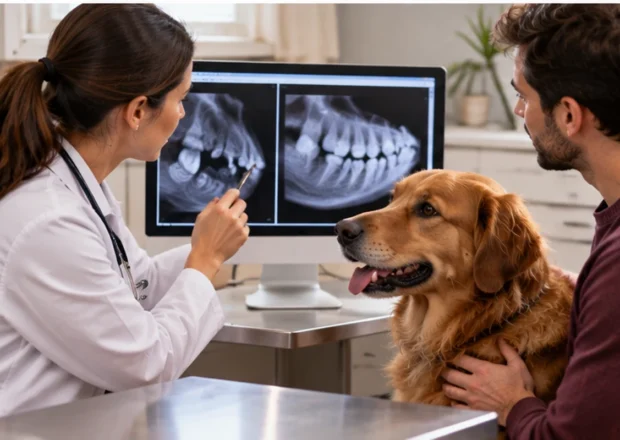
Radiographs use X-ray energy to create images of tissues with different densities. They are particularly useful for evaluating many bones, certain chest structures and some abdominal patterns. A veterinarian might order them after a suspected fracture, difficulty breathing, abdominal distension or concern about a swallowed object. In these cases, the clinical question drives image selection. The pet’s symptoms and examination usually determine whether radiographs are appropriate before cost enters the conversation.
An X-ray is not a universal camera that identifies every illness. Some objects are hard to see, and soft-tissue detail may be limited compared with ultrasound, CT or other testing. Certain conditions need serial images, contrast studies or follow-up work even after the initial radiograph appears unremarkable. Specialist interpretation may also change the next step. VCA’s imaging guidance discusses how radiography, ultrasound and other tools complement one another. Ask the veterinarian which diagnosis the proposed images are intended to help confirm or exclude, and what decision depends on the result.
Number of views and body region
A study may require several positions or “views” of the same region because a two-dimensional image can hide information that becomes clearer from another angle. A chest study, limb series and abdominal study are not identical products. Some clinics quote by region and expected view count; others bundle multiple images in a single procedure. If the first set is technically inadequate because a pet moves or an area is hard to position, new images may sometimes be necessary.
Ask whether the estimate includes two or three views, whether additional images are billed separately and what happens if the first result points to another body region. When comparing two quotes, ensure that each clinic is proposing the same study rather than the same word, “X-ray.” A lower amount for one view cannot fairly be presented as less expensive than a more comprehensive series. The relevant question is not how many pictures a clinic sells but what the veterinarian needs to answer the medical question safely.
Sedation, restraint and comfort
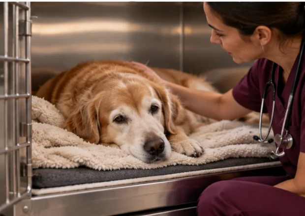
Some animals tolerate careful positioning while awake. Others may be frightened, painful, difficult to hold still or unable to maintain a necessary posture. A veterinary team may recommend sedation or anesthesia when appropriate to obtain diagnostic images safely and minimize distress. These services can add charges for medication, monitoring, a pre-sedation assessment and recovery observation. Not every pet needs them, and not every possible image can be captured adequately without them.
For a dog with a painful injury, forcing a limb into position may worsen stress and make the image less useful. For an anxious cat, a clinician may weigh the risks and benefits of chemical restraint rather than repeated physical attempts. Age, concurrent disease and emergency stability also influence the plan. Ask whether sedation is expected, what monitoring is included, who supervises recovery and whether the estimate covers a contingency if additional intervention is required. A low advertised imaging fee that excludes all sedation-related care may not represent the final amount.
Interpretation and the possible specialist fee
Taking the image and interpreting it are related but separate tasks. A general-practice veterinarian may review radiographs directly, while a more complicated study may be sent to a veterinary radiologist for a formal report. Emergency hospitals and referral clinics can also involve clinicians with different expertise. A specialist interpretation may add a charge, affect timing and offer further detail when findings are subtle or when surgery is being considered. It should not be assumed that every imaging invoice includes the same level of review.
When the clinic gives you the estimate, ask whether interpretation is included, whether a written report will be provided, and whether digital images can be transferred to your primary veterinarian or referral center. Access to the images can reduce confusion if your pet later needs additional care, but not every receiving clinic will regard old images as a substitute for new ones. If the diagnosis remains uncertain, ask what further test might be recommended, why it would answer a different question and how the cost would be discussed before it is performed.
A sample invoice that is explicitly hypothetical
Here is a teaching example, not a survey or real clinic quote. Imagine an outpatient hospital estimates $180 for the radiographic study, $70 for the office examination, $45 for imaging interpretation and $100 for sedation plus monitoring. The full modeled total is $395, not the $180 headline imaging line. In a second purely hypothetical case, the patient does not need sedation and interpretation is included in the study price, producing a smaller total. Neither scenario predicts what any U.S. provider charges today.
Use this arithmetic to compare the scope of estimates, not to select a clinic from fictional prices. An actual invoice may also include emergency triage, laboratory work, medication, supplies, taxes where applicable and follow-up visits. Ask the hospital to distinguish expected charges from optional or conditional steps. If it gives a range, ask what clinical finding would push the estimate toward the upper end. A written explanation of what is and is not included often prevents more confusion than a vague promise that imaging is inexpensive.
When X-rays lead to ultrasound, CT or surgery
Consider a dog vomiting after chewing a fabric toy. A veterinarian may recommend abdominal radiographs because some obstruction patterns can be visible, but a soft material may not appear as a sharply outlined object. Merck’s discussion of gastrointestinal obstruction explains the role of radiographs and ultrasound in diagnosing a potentially serious blockage. If the images do not resolve the clinical concern, more testing or surgery may be necessary. That progression does not mean the first test was automatically pointless.
Likewise, radiographs of a limb can reveal certain fractures while leaving questions about ligaments or soft tissues unresolved. Advanced imaging may be available through specialty facilities and carries a separate cost structure. Ask what the initial study is expected to contribute, what result would justify a second study, and whether the clinic can provide an updated estimate at each decision point. In an emergency, medical stabilization may take priority over separating the bill into neat elective steps; safety still comes before comparison shopping.
Insurance implications: the word X-ray is not enough
Accident-and-illness pet insurance often lists diagnostic imaging among potentially covered services, but approval depends on the reason for the study and the rest of the policy. A radiograph ordered to investigate a new eligible injury may receive different treatment from imaging related to a condition documented before coverage or performed during an applicable waiting period. An exam charge, sedation or interpretation could be covered, limited or excluded according to policy terms. A wellness arrangement may have entirely separate rules.
Record the veterinarian’s reason for the test and keep the complete clinical note, imaging report and itemized invoice. If your plan reimburses eligible expenses after a deductible, calculate from the eligible amount rather than assuming a percentage of the whole checkout total. Annual limits and previously paid claims can further restrict the payment. Contact the insurer for documentation requirements, but do not delay necessary imaging while seeking a preliminary coverage opinion. Insurers may request historical records even when the pet has no microchip or formal prior diagnosis.
How to ask for an estimate respectfully and clearly
You can ask the clinic: Which body area is being imaged? How many views do you expect? Is the physical examination included? Will my pet need sedation or monitoring? Who interprets the images? Is a referral or radiologist review included? What would make the total rise? When would I be asked to approve an additional test? These questions let the clinician explain both the medical value and the financial scope without suggesting that the lowest priced procedure is always adequate.
For planned nonurgent imaging, compare written estimates from facilities that can perform the medically recommended study. For emergencies, ask which decisions cannot safely wait and whether any available payment assistance or staged diagnostics would be medically reasonable. AAHA encourages owners to prepare for veterinary expenses and discuss financial planning rather than treating payment problems as taboo. Save the invoice and report for future follow-up, because the medical record can be at least as useful as the price when your pet returns with another concern.
How to compare imaging estimates without choosing the wrong test
Start with the clinical question rather than an advertised scan price. If one veterinarian recommends two abdominal views and another recommends a larger series with sedation, the quotations are not equivalent. Ask the clinician to describe what each view contributes, whether additional images might be needed and how likely a repeat study is to affect the decision. If an estimate is for a specialist referral, clarify whether the receiving team will re-examine the pet and charge separately for its own interpretation.
For nonurgent appointments, ask whether copies of digital images and a written report are included. If a previous clinic recently took images, the new veterinarian may be able to review them, though repeat imaging may still be medically justified when symptoms have changed or the older study lacks the required views. An owner cannot insist that old images provide the answer to a new question; instead, discuss what extra information a fresh study is expected to provide.
When an animal is in severe distress, use the estimate discussion to understand the next decision, not to postpone stabilization. The financial trade-off of a small saving is different when a delay could allow an obstruction or breathing problem to worsen. Clear written expectations, permission for extra charges and an agreed contact number help the care team communicate promptly if the first study changes the plan.
Research sources
- VCA: Radiographs for Dogs
- VCA: Radiographs for Cats
- VCA Animal Specialty Group: Diagnostic Imaging
- Merck: Gastrointestinal Obstruction Imaging Context
- AAHA: Preparing for Pet Healthcare Costs
Research checked October 9, 2026. Educational veterinary articles do not diagnose a pet or replace individual veterinary care. Any financial calculation is hypothetical; insurance coverage is determined by the issued policy and claim facts.